This MRI is most consistent with which of the following diagnoses?
|
| ||||||||||||||||||||||||||||||||||||||||||||||||||||||||||||||||||||||||||||||||||||||||||||||||||||||||||||||||
Neurocutaneous Syndromes 05Topic: ImagingCreated on Friday, September 21 2007 by jdmiles Last modified on Friday, September 21 2007.
This MRI is most consistent with which of the following diagnoses? A) Tuberous sclerosis B) Ehlers-Danlos syndrome C) Neurofibromatosis type 1 D) Neurofibromatosis type 2 E) Von Hippel-Lindau disease
This question was last modified on September 21, 2007.
ANSWERS AND EXPLANATIONSA) Tuberous sclerosis
| ||||||||||||||||||||||||||||||||||||||||||||||||||||||||||||||||||||||||||||||||||||||||||||||||||||||||||||||||
 |  |  | 
|  |  |
| Please log in if you want to rate questions. | |||||
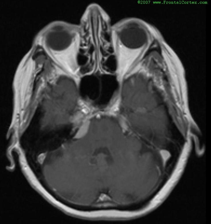
The MRI shows bilateral acoustic neuromas. This is a classic feature of neurofibromatosis type 2 (NF2).
Ehlers-Danlos syndrome (EDS) does not typically present with acoustic neuromas. Radiographically visible lesions typically associated wtih EDS include large, sometimes multiple aneurysms.
(See References) | | |
| | |
| Please log in if you want to rate questions. | |||||
The MRI shows bilateral acoustic neuromas. This is a classic feature of neurofibromatosis type 2 (NF2).
Neurofibromatosis type 1 (NF1, von Recklinghausen disease) does not typically present with acoustic neuromas. Typical findings in NF1 include cafe au lait spots, axillary freckling, palpable neurofibromas, Lisch nodules, optic glioma, and bone lesions.
(See References) | | |
| | |
| Please log in if you want to rate questions. | |||||
| | |
| | |
| Please log in if you want to rate questions. | |||||
The MRI shows bilateral acoustic neuromas. This is a classic feature of neurofibromatosis type 2 (NF2).
Von Hippel-Lindau disease (vHL) does not typically present with acoustic neuromas. Radiographically visible lesions typically associated wtih vHL include hemangioblastomas, especially in the cerebellum and retina.
(See References) | | |
| | |
| Please log in if you want to rate questions. | |||||
| 1. Rowland, L.P. (Ed) (2000). Merritt's Neurology, 10th Edition. Lippincott Williams & Wilkins, Philadelphia. | |
| 2. Fenichel, G.M. (2005). Clinical Pediatric Neurology, 5th ed. Elsevier, Philadelphia. | |
| 3. Santos, C.C., Miller, V.S., and Roach, E.S. (2004). Neurocutaneous syndromes. In Bradley, W.G., Daroff, R.B., Fenichel, G.M., and Jankovic, J. (Eds.). Neurology in Clinical Practice, Fourth Edition. Butterworth Heinemann, Philadelphia, pp. 1867-1900. | |
| 4. Victor, M., and Ropper, A.H. (2001). Adams and Victor's Principles of Neurology, 7th Edition. McGraw-Hill, New York. | |
| 5. Prayson, R.A., and Goldblum, J.R. (Eds.) (2005). Neuropathology. Elsevier Churchill Livingstone, Philadelphia. |
| | |
| | |
| Please log in if you want to rate questions. | |||||
FrontalCortex.com -- Neurology Review Questions -- Neurology Boards -- Board Review -- Residency Inservice Training Exam -- RITE Exam Review
log in to FrontalCortex.com
New to FrontalCortex?
|

![]()
![]()

| | We comply with the HONcode standard for trustworthy health information: verify here. |
Share this page:
 |  |
 |
 |
 |
 |
 |
 |
 |
 |
 |
 |
 |
|