Vascular Malformations 05
Topic: Pathology
Created on Thursday, November 29 2007 by jdmiles
Last modified on Thursday, November 29 2007.
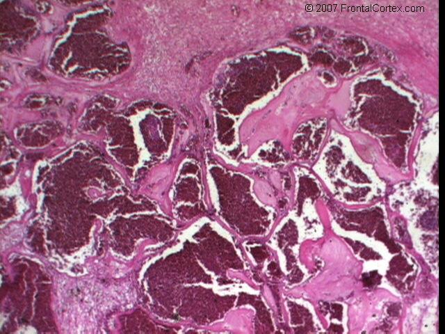
Which of the following statements about the type of lesion seen in this photomicrograph is most accurate?
This question was created on November 29, 2007 by jdmiles.
This question was last modified on November 29, 2007.
ANSWERS AND EXPLANATIONS
A) this is a capillary telangiectasia
This answer is incorrect.
This photomicrograph shows a cavernous angioma. Cavernous angiomas are most often seen in young adults, and 1/3 of cases present with focal seizures. There is an approximately 1% annual risk of hemorrhage with this type of lesion. Microscopically, cavernous angiomas appear as clusters of dilated, thin-walled blood vessels with no brain tissue in between. Unlike AVMs, there is no arterial component. (
See References)
|
 |  |  | 
|  |  |
| Please log in if you want to rate questions. |
B) this is an arteriovenous malformation
This answer is incorrect.
This photomicrograph shows a cavernous angioma. Cavernous angiomas are most often seen in young adults, and 1/3 of cases present with focal seizures. There is an approximately 1% annual risk of hemorrhage with this type of lesion. Microscopically, cavernous angiomas appear as clusters of dilated, thin-walled blood vessels with no brain tissue in between. Unlike AVMs, there is no arterial component. (
See References)
|
| | |
| | |
| Please log in if you want to rate questions. |
C) this type of lesion is seen most often in young adults
This answer is correct.
This photomicrograph shows a cavernous angioma. Cavernous angiomas are most often seen in young adults, and 1/3 of cases present with focal seizures. There is an approximately 1% annual risk of hemorrhage with this type of lesion. Microscopically, cavernous angiomas appear as clusters of dilated, thin-walled blood vessels with no brain tissue in between. Unlike AVMs, there is no arterial component. (
See References)
|
| | |
| | |
| Please log in if you want to rate questions. |
D) lesions of this type are dilated veins of the superficial or subcortical vasculature
This answer is incorrect.
This photomicrograph shows a cavernous angioma. Cavernous angiomas are most often seen in young adults, and 1/3 of cases present with focal seizures. There is an approximately 1% annual risk of hemorrhage with this type of lesion. Microscopically, cavernous angiomas appear as clusters of dilated, thin-walled blood vessels with no brain tissue in between. Unlike AVMs, there is no arterial component. (
See References)
|
| | |
| | |
| Please log in if you want to rate questions. |
E) this type of lesion is associated with a 2%-4% annual risk of acute hemorrhage
This answer is incorrect.
This photomicrograph shows a cavernous angioma. Cavernous angiomas are most often seen in young adults, and 1/3 of cases present with focal seizures. There is an approximately 1% annual risk of hemorrhage with this type of lesion. Microscopically, cavernous angiomas appear as clusters of dilated, thin-walled blood vessels with no brain tissue in between. Unlike AVMs, there is no arterial component. (
See References)
|
| | |
| | |
| Please log in if you want to rate questions. |
References:
| 1. Prayson, R.A., and Goldblum, J.R. (Eds.) (2005). Neuropathology. Elsevier Churchill Livingstone, Philadelphia. (ISBN:0443066582)
| Advertising:
|
|
| | |
| | |
| Please log in if you want to rate questions. |
FrontalCortex.com -- Neurology Review Questions -- Neurology Boards -- Board Review -- Residency Inservice Training Exam -- RITE Exam Review
pathology
Vascular Malformations 05
Question ID: 11290703
Question written by J. Douglas Miles, (C) 2006-2009, all rights reserved.
Created: 11/29/2007
Modified: 11/29/2007
Estimated Permutations: 297000