FrontalCortex, Inc. is a non-profit corporation dedicated to neurology education.

Optic Pathway Gliomas 01

Topic: Imaging

Created on Sunday, October 12 2008 by jdmiles

Last modified on Sunday, October 12 2008.


Regarding the tumor seen in this image, which of the following statements is most accurate?
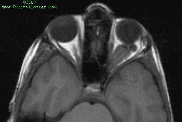
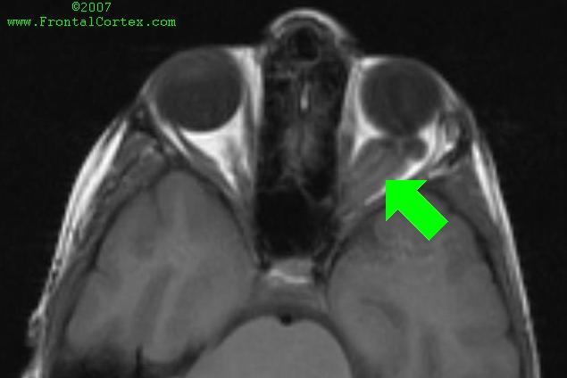
 
        A) Tumors of this kind are usually diffuse astrocytomas
 
        B) Tumors of this kind are usually pilocytic astrocytomas.
 
        C) Tumors of this kind are seen more frequently in patients with von Hippel-Lindau disease
 
        D) Tumors of this kind are usually subependymal giant cell astrocytomas
 
        E) Tumors of this kind are usually pleomorphic xanthoastrocytomas


Back to the question = Go back to the top of the page.
See another question like this one = Reload a different version of this question ().
Click here for a random question = Load a random question from the database.
Clone this question = Use this question as a template to create a totally NEW question.
Rate this question = Enter detailed rating for this question!
Average user rating for this question = 4 = How users like you have rated this question.
This question was created on October 12, 2008 by jdmiles.
This question was last modified on October 12, 2008.


ANSWERS AND EXPLANATIONS


A) Tumors of this kind are usually diffuse astrocytomas

This answer is incorrect.


The MRI shows an optic glioma behind the left eye. Optic pathway gliomas are a common finding in neurofibromatosis type 1 (NF1). Optic pathway gliomas are usually pilocytic astrocytomas.  (See References)

Back to the questionSee another question like this oneClick here for a random questionClone this question Rate this questionAverage user rating for this question = 4
Please log in if you want to rate questions.


B) Tumors of this kind are usually pilocytic astrocytomas.

This answer is correct.


The MRI shows an optic glioma behind the left eye. Optic pathway gliomas are a common finding in neurofibromatosis type 1 (NF1). Optic pathway gliomas are usually pilocytic astrocytomas.  (See References)

Back to the questionSee another question like this oneClick here for a random questionClone this question Rate this questionAverage user rating for this question = 4
Please log in if you want to rate questions.


C) Tumors of this kind are seen more frequently in patients with von Hippel-Lindau disease

This answer is incorrect.


The MRI shows an optic glioma behind the left eye. Optic pathway gliomas are a common finding in neurofibromatosis type 1 (NF1). Optic pathway gliomas are usually pilocytic astrocytomas.
Von Hippel-Lindau disease (vHL) does not typically present with an optic glioma. Radiographically visible lesions typically associated wtih vHL include hemangioblastomas, especially in the cerebellum and retina.  (See References)

Back to the questionSee another question like this oneClick here for a random questionClone this question Rate this questionAverage user rating for this question = 4
Please log in if you want to rate questions.


D) Tumors of this kind are usually subependymal giant cell astrocytomas

This answer is incorrect.


The MRI shows an optic glioma behind the left eye. Optic pathway gliomas are a common finding in neurofibromatosis type 1 (NF1). Optic pathway gliomas are usually pilocytic astrocytomas.  (See References)

Back to the questionSee another question like this oneClick here for a random questionClone this question Rate this questionAverage user rating for this question = 4
Please log in if you want to rate questions.


E) Tumors of this kind are usually pleomorphic xanthoastrocytomas

This answer is incorrect.


The MRI shows an optic glioma behind the left eye. Optic pathway gliomas are a common finding in neurofibromatosis type 1 (NF1). Optic pathway gliomas are usually pilocytic astrocytomas.  (See References)

Back to the questionSee another question like this oneClick here for a random questionClone this question Rate this questionAverage user rating for this question = 4
Please log in if you want to rate questions.


References:

1. Prayson, R.A., and Goldblum, J.R. (Eds.) (2005). Neuropathology. Elsevier Churchill Livingstone, Philadelphia. (ISBN:0443066582)Advertising:
2. Victor, M., and Ropper, A.H. (2001). Adams and Victor's Principles of Neurology, 7th Edition. McGraw-Hill, New York. (ISBN:0070674973)Advertising:
3. Santos, C.C., Miller, V.S., and Roach, E.S. (2004). Neurocutaneous syndromes. In Bradley, W.G., Daroff, R.B., Fenichel, G.M., and Jankovic, J. (Eds.). Neurology in Clinical Practice, Fourth Edition. Butterworth Heinemann, Philadelphia, pp. 1867-1900 (ISBN:0750674695).Advertising:
4. Rowland, L.P. (Ed) (2000). Merritt's Neurology, 10th Edition. Lippincott Williams & Wilkins, Philadelphia. (ISBN:0683304747)Advertising:
5. Fenichel, G.M. (2005). Clinical Pediatric Neurology, 5th ed. Elsevier, Philadelphia. (ISBN:1416001697)Advertising:
Back to the questionSee another question like this oneClick here for a random questionClone this question Rate this questionAverage user rating for this question = 4
Please log in if you want to rate questions.


FrontalCortex.com -- Neurology Review Questions -- Neurology Boards -- Board Review -- Residency Inservice Training Exam -- RITE Exam Review
imaging
Optic Pathway Gliomas 01
Question ID: 101208162
Question written by J. Douglas Miles, (C) 2006-2009, all rights reserved.
Created: 10/12/2008
Modified: 10/12/2008
Estimated Permutations: 8400

User Comments About This Question:

0 user entries
Please log in if you'd like to add a comment.